Workshops
For the 2026 edition of NESS, you’ll have the opportunity to choose from multiple workshops, each designed to cover different aspects of neural engineering. We’ve curated a diverse selection to ensure a broad and hands-on learning experience, with workshops requiring various backgrounds and skill sets.
During registration, you’ll be asked to rank the workshops based on your interests and expertise. Once the application process is complete, we will assign workshops to selected participants, aiming to match everyone with their top preferences.
Please note: You won’t be able to attend all workshops, as it’s not possible to fit them all into a single week. Choose wisely to make the most of your experience!
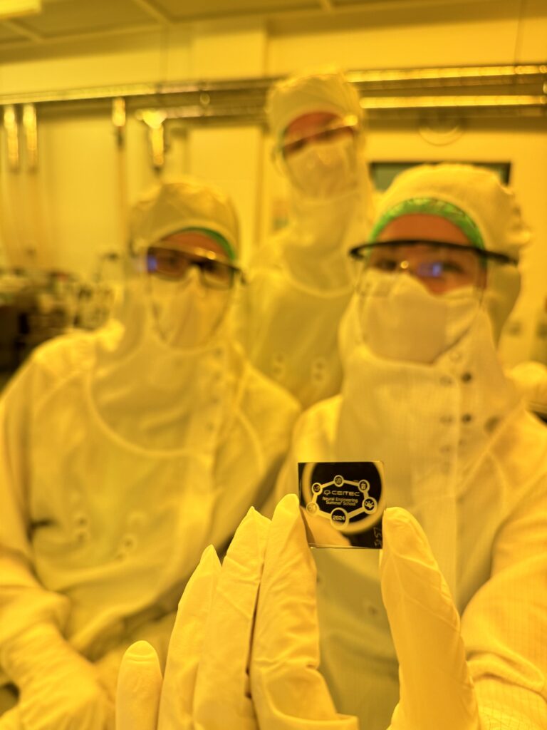
W1: Introduction to Photolithography: Patterning a Thin-Film Metal Layer
Discover the fundamentals of photolithography, a cornerstone technique in microfabrication used to create intricate patterns on thin films for modern electronic devices. In this hands-on exercise, you will step into a cleanroom environment and participate in patterning a thin-film metal layer to produce a small, personalized logo of our summer school—yours to take home.
You will learn about the key steps involved in the process:
- Photoresist Coating: Apply a light-sensitive material (photoresist) onto the metal-coated substrate.
- UV Exposure: Use a photomask and UV light to selectively expose the photoresist, transferring a specific pattern onto the metal layer.
- Development: Chemically develop the photoresist, revealing the exposed areas while protecting others.
Etching: Remove the unprotected metal areas to create the final patterned structure.
This exercise offers a unique opportunity to experience microfabrication up close and gain insight into the processes behind creating advanced neuroelectronic devices. Whether you are looking to explore the world of cleanroom techniques or simply want a hands-on introduction to how cutting-edge technologies are made, this session provides an accessible and engaging experience.
Duration: ½ day
W2: Noninvasive Electrical Stimulation of Peripheral Nerves
Transcutaneous electrical stimulation of peripheral nerves is a noninvasive way to access the human nervous system, and is used in routine diagnostic procedures as well as bioelectronic medicine to treat conditions like chronic pain, inflammatory disorders, psychiatric conditions, and movement disorders. The technique is also vital in many rehabilitation and physiotherapy/sports medicine applications. In this activity, you will perform practical nerve stimulation experiments on each other, demonstrating key biophysical principles which govern how this kind of stimulation works, and dictating its scope of limitations.
Stimulation of sensory fibers: Fast alpha-type fibers transmit sensory stimuli from the periphery to the central nervous system, we will learn how to selectively stimulate them.
Stimulation of motor fibers – muscle actuation: Fast alpha-type motor fibers actuate our skeletal muscles, and this kind of stimulation is common in physiotherapy. We will learn how to selectively stimulate different muscles.
The strength-duration relation: We will demonstrate a core biophysical concept governing electrical stimulation and used to differentiate different nerve types, how current thresholds required to achieve stimulation vary with the length of the stimulation pulse.
Supraphysiological frequencies – temporal summation effects: You will see how frequencies into the range >1000 Hz can stimulate via delivering successive subthreshold stimulation pulses, to achieve unique patterns of stimulation of sensory and motor fibers.
This exercise is very hands-on and practical, and theoretical concepts are presented during the experiments themselves. Participants will take turns acting as “experimenter” and “subject”, with the goal of understanding important considerations in designing bias-free experiments.
Duration: ½ day
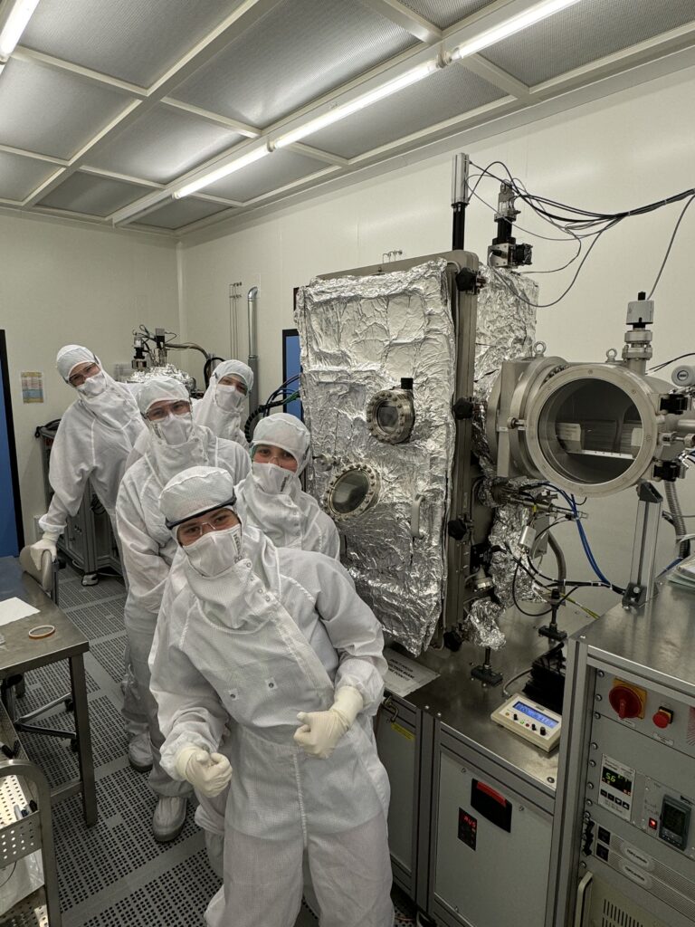
W3: Microfabrication of an Ultrathin Stimulation Electrode Array for Peripheral Nerve Applications
This two-day hands-on exercise is designed for participants who want a deeper dive into microfabrication and thin-film technology used in neuroengineering. The workshop provides practical exposure to key fabrication techniques commonly employed in the development of ultrathin stimulation electrode arrays.
Each participant will fabricate their own ultrathin stimulation electrode array by working through selected steps of a representative fabrication workflow from initial material deposition to final release and inspection. The process combines cleanroom-based microfabrication with complementary general laboratory techniques, reflecting realistic research workflows.
Participants will gain hands-on experience with:
- Thin-film deposition: Applying metal and insulating layers using selected deposition techniques commonly used in microfabrication.
- Patterning and etching: Structuring metal and dielectric layers using photolithography, followed by wet and dry etching processes.
- Complementary fabrication techniques: Demonstrations of laser-based micromachining and electrodeposition, highlighting approaches used for rapid prototyping, post-processing, and functional surface modification.
The session will conclude with a short device characterization step, where participants will assess the quality of the fabricated structures. For those interested, a follow-up session will be available to explore testing of microfabricated stimulation electrodes in a biological model using an in vivo locust preparation.
Duration: 2 days
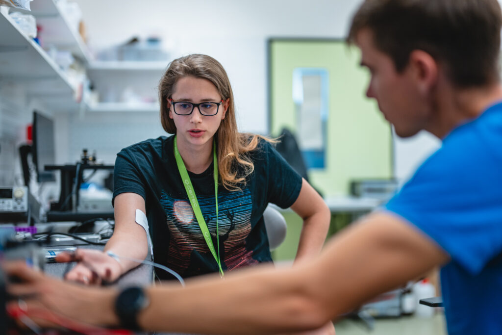
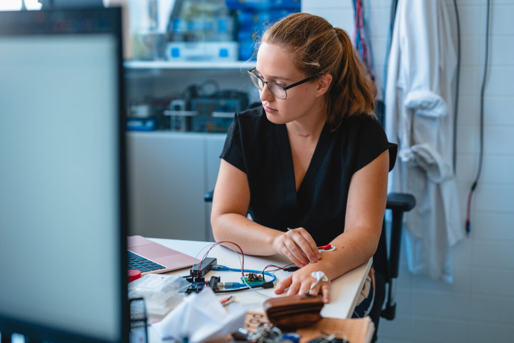
W4: DIY EMG Amplifier
Explore the fundamentals of bioelectrical signal acquisition by building your own EMG (electromyography) amplifier from scratch. In this hands-on session, you will assemble a working EMG amplifier by soldering discrete components onto a printed circuit board and then test it on your own muscles to observe and analyze real-time EMG signals.
You will learn about the key steps involved in the process:
- Circuit Assembly: Solder electronic components onto a PCB to construct a functional EMG amplifier.
- Signal Acquisition: Properly place electrodes to measure muscle activity and understand factors influencing signal quality.
- Arduino Programming: Write a script to display and filter EMG data for visualization and analysis.
- Amplifier Fundamentals: Understand the basic principles of amplifier circuits and their role in bioelectrical signal processing.
- Electrode Selection & Placement: Learn about different electrode types and how placement affects signal strength and clarity.
This workshop offers a hands-on introduction to bio-signal acquisition and practical electronics, providing you with essential skills in circuit design, soldering, and signal processing. Whether you are new to neural engineering or looking to deepen your understanding of EMG systems, this session will give you an engaging and interactive experience with real-world applications.
Duration: ½ day
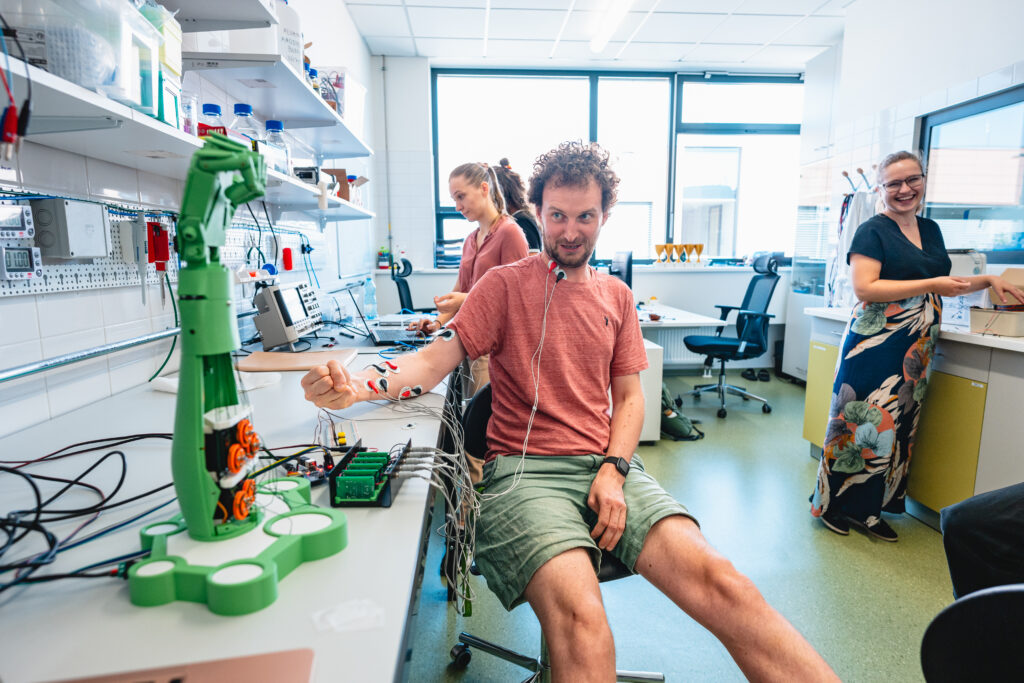
W5: Introduction to the Brain Machine Interfaces
Step into the world of Brain-Machine Interfaces (BMI) by leveraging EMG signals to control both machines and human muscles. In this advanced hands-on session, you will use multiple DIY EMG amplifiers to decode muscle activity associated with individual finger movements. You will then apply this knowledge to control a robotic arm and electrically stimulate another person’s muscles, effectively transferring movement from one person to another.
You will learn about the key steps involved in the process:
- Advanced EMG Signal Acquisition & Processing: Record, filter, and classify EMG signals from different muscles to decipher movement intentions.
- Robotic Control with Arduino: Program and interface a robotic arm to respond to EMG commands.
- Electrical Muscle Stimulation (EMS): Learn the principles of neuromuscular stimulation and how to trigger muscle contractions in another person based on EMG signals.
- Electrode Selection & Placement: Explore how different electrode types and positioning affect signal quality and stimulation targeting.
- BMI Applications: Gain insights into real-world applications of EMG-based control systems in prosthetics, rehabilitation, and assistive technologies.
This session is designed for students with prior experience in programming and signal processing who want to explore cutting-edge neural engineering techniques. By the end of the class, you will have hands-on experience in decoding bioelectrical signals and using them to control both machines and human muscles—providing a deeper understanding of the principles behind modern Brain-Machine Interfaces.
Duration: 1 day
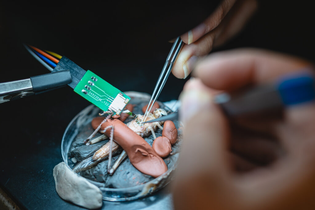
W6: Neural Stimulation and Recording with Invertebrates
Extracellular recording and stimulation is a fundamental electrophysiology technique for studying nervous system physiology and also is used for prototyping new neural interface technologies. Traditionally, nearly all work is done using rodents like mice and rats. However, this represents a significant financial and ethical burden on research teams, and we strongly advocate the use of invertebrate models like annelids and insects as a suitable replacement for mammalian models. In this experimental module, you will learn fundamental electrophysiology techniques using two insect models: the locust and the cockroach.
Extracellular neural recording: You will learn how to use a multichannel biopotential recording amplifier to collect electrical activity from different regions of the insect nervous system, and how to filter and sort the data to understand firing behaviour and electrical oscillations of neural circuits.
Microstimulation and electrophygraphy: We will apply a fundamental technique in studying evoked activity by using microelectrodes to stimulate a motor nerve in the locust and recording electromyography in the leg muscle.
Supraphysiological frequencies and nerve block: You will see how frequencies into the range >1000 Hz can stimulate via delivering successive subthreshold stimulation pulses, to achieve selective stimulation or neural blocking effects.
This exercise is hands-on and practical, and theoretical concepts are presented during the experiments themselves. Participants will perform most experiments themselves. Dissection work is required, involving sharp tools like scissors and scalpels, as well as microneedle electrodes.
Duration: ½ day
W7: In Vivo Testing of Ultrathin Neurostimulation Devices in a Locust Model
In this (follow-up) session, participants will test their microfabricated neurostimulation devices in a biological model, gaining hands-on experience with in vivo neurostimulation experiments. The locust model provides a robust and accessible system for studying nerve stimulation due to its well-defined peripheral nervous system. Specifically, participants will target the N5 nerve, which controls the locust’s jumping leg. This nerve is used in neuroengineering research as it offers a clear and observable motor response to stimulation.
Key steps in this exercise will include:
- Surgical preparation: Exposing the N5 nerve by carefully dissecting the surrounding tissue while ensuring the nerve remains intact and functional.
- Device placement and implantation: Positioning and securing the ultrathin electrode array or wireless neurostimulator onto the nerve.
- Neurostimulation experiments: Recording the strength-duration relationship, a key parameter that describes the relationship between stimulation pulse intensity and duration required to activate the nerve.
This exercise is open to participants from the Microfabrication of an Ultrathin Stimulation Electrode Array for Peripheral Nerve Applications session, allowing them to test their own devices. Those who did not participate in the fabrication process can still join and work with pre-made devices.
This lab provides an opportunity to bridge microfabrication and neuroengineering, demonstrating how thin-film stimulation devices interact with living tissue and how stimulation parameters influence nerve activation.
Duration: ½ day
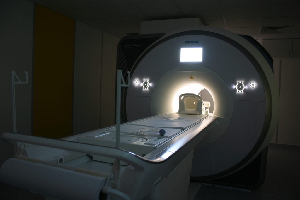
W8: What and How Can MRI Measure?
Magnetic Resonance Imaging (MRI) is an imaging technique that uses a strong magnetic field to create detailed images of the internal structures of the body. MRI does not use ionizing radiation, making it a safe method for patients. MRI can measure a wide range of parameters that allow us to study not only the structure of tissues, but also their functional behaviour and composition.
In this activity you will measure MRI data, test how the parameters of the measurement sequence affect the resulting image and explore how the imaging process can be affected by the presence of various sources of noise.
MR safety and measurements: You will learn what needs to be done to measure and why. You will try to measure your own MRI data.
Measurement parameters: You will test the influence of scan parameters on the result and the quality of the MR image.
Artefacts: You will learn and explore how different sources of noise can affect the MR image or other measured physiological signals (e.g. ECG).
This exercise involves data collection and experimental testing and provides hands-on experience with a whole-body MR scanner. You will learn how to control the MR scanner, get the practical background to measure good quality data and understand what kind of information MR scans can provide.
Duration: ½ day
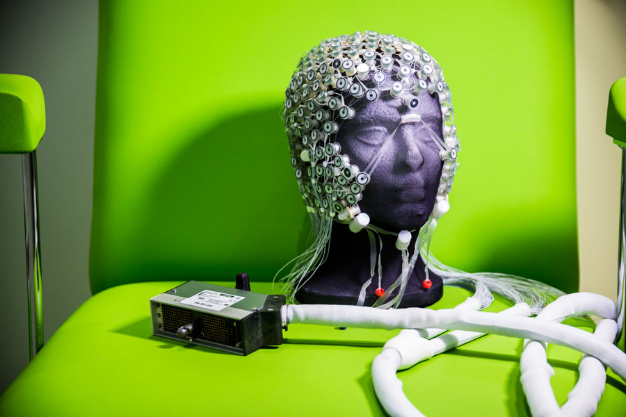
W9: Electroencephalography – Non-invasive measurement of brain activity
It has been more than a hundred years since Hans Berger acquired the first electroencephalogram, a non-invasive recording of the electrical activity of the human brain, and described the so-called alpha waves. The technology has come a long way since then, but the basics remain the same. Electroencephalography can track brain activity with very good temporal and reasonable spatial resolution, and it helps to better understand the physiology of the human brain.
In this course you will record a task-based EEG of each other, learn how to process the data and how to interpret the results.
EEG Measurement: You will learn how to set up an EEG experiment and record good quality data. We will focus on the open/closed eye block design experiment.
EEG processing: The EEG data you record needs to be analysed, we will learn how to do this properly. We will focus on alpha oscillatory activity in the occipital lobe (visual cortex).
Brain-computer interface: Why not process the data instantly, online, during the measurement and try to follow the fluctuations of the alpha activity online?
Movement: All these new experiences will be used to prepare a simple task where you will follow the activity over the motor cortex, process the data online and try to judge which arm is performing the movement.
This exercise is very practical and hands-on. It involves data collection, post-hoc analysis and also on-line data processing. Participants will be involved in all these steps.
Duration: 1 day
W10: Transcranial Magnetic Stimulation
Transcranial magnetic stimulation (TMS) is a safe non-invasive method that is based on the Faraday’s principle of electromagnetic induction. The special electromagnetic coil is placed over the head and through rapid changes of a magnetic field induces an electric current in the region of the brain under the coil. The induction of this secondary electric current at sufficient intensity can directly activate cortical neurons at a depth of 1.5-2 cm beneath the scalp This technique can be used in research for measurement of neural excitability or in clinical practice for treatment of psychiatric and neurological disorders. During this activity, you will apply TMS to each other’s motor cortex to induce movement in your limbs and measure the motor threshold. You will also utilize a specialized neuronavigation system to achieve precise coil placement over the head.
Identifying Neural Coordinates: You will learn to use MNI coordinates for brain stimulation and apply a MATLAB script to preprocess MRI scans to determine motor cortex coordinates.
Preparation of a 3D Model of the Head and Brain: You will learn how to create a 3D model of the head and brain using BrainSight neuronavigation software.
Navigation of the Coil and Stimulation: You will learn how to manipulate the TMS device and use neuronavigation for precise coil placement.
Measurement of Motor Threshold: You will see how magnetic pulses can directly activate neurons in your motor cortex and induce movement in your limbs. You will learn how to use electromyography to measure muscle activations.
This exercise is very hands-on and practical, and theoretical concepts are presented during the experiments themselves. Participants will take turns acting as “experimenter” and “subject”. All participants will undergo a measurement inside MR scanner.
Duration: ½ day
W11: Dynamical Models of Brain Activity
Making sense of the experimental observations in neuroscience implies understanding how brain structure, function and dynamics relate to each other. Models of different scopes and scales can not only help us provide mechanistic explanations but also can be incorporated in inference workflows. In this one-day exercise we will build and explore models of the EEG and TMS experiments performed in other sessions (participation not a requirement, but beneficial). In particular, the participants will:
Build and run the model: we will learn which building blocks are used to construct the model of the selected experimental paradigm, and compare the simulated readout with the empirical data
Understand the model: by perturbing the parameters in a systematic way, we will learn about the sensitivity of the model output to changes in the parameter values
Tinker: we will modify the model to explore additional scenarios: different stimulation site, EEG readout for the TMS stimulus, eyes-opened vs eyes-closed for the spontaneous activity.
The session is organized as a guided tour through the modelling workflows, with the theoretical background being provided as we go. The participants will run and evaluate the models themselves, and by the end of the session will have a basic understanding how the computational and experimental work can interact. No prior experience with numerical computation is required, basic familiarity with Python or Matlab is expected.
Duration: 1 day
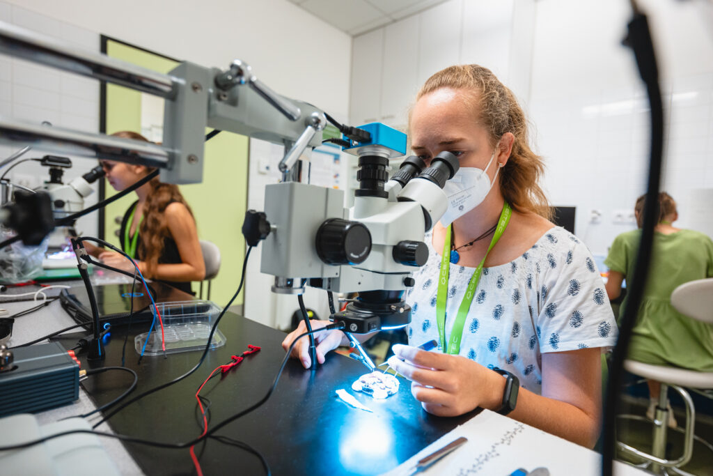
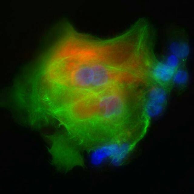
W12: Fundamental Neuronal Cell Culture Techniques
Developing in-vitro cell culture models of neural tissue is becoming an increasingly indispensable aspect of the field of Neural Engineering. These techniques can be used to test and refine devices and study neural interfaces, as well as to provide preclinical evidence for their effectiveness in disease models. Learning the fundamentals of these techniques and understanding how to work in sterile conditions to culture and manipulate different types of cell preparations is a valuable skill in such an interdisciplinary field.
Scope of Activity:
- Primary neuronal cell isolation from chicken embryos
- Cell manipulation and maintenance techniques using a glial cell line
- Immunohistochemical staining for confocal microscopy
Duration: 2 and ½ Day
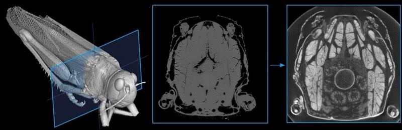
W13: X-ray Computed Microtomography
Can we visualise the brain and nerves using X-rays? X-ray computed microtomography (microCT) is a non-destructive imaging technique that can serve as a powerful tool in biological research. In this workshop, we will explore the possibilities and limitations of microCT for neural imaging by examining biological samples ex vivo.
Scope of Activity:
- MicroCT Scanning: First, we will prepare samples for microCT scanning. Participants will learn how to mount specimens so they can rotate 360° during acquisition and how to select appropriate scanning parameters. We will then launch a scan and evaluate the results.
- Phase Contrast or Staining? Next, we will discuss strategies for increasing soft-tissue contrast, including phase-contrast imaging and contrast-agent staining. A second scan will be performed, and the two datasets will be compared to determine which approach provides better results for analysis.
- Creation of a 3D Model: Finally, we will introduce basic image analysis concepts and demonstrate approaches for creating and exploring 3D reconstructions.
No previous experience is required. In this half-day workshop, we will cover the fundamentals of X-ray microCT imaging and discuss its possibilities and challenges.
Duration: ½ day